


Fever Package (includes Malaria & Typhoid tests) Old

For men & women
Earliest reports in

12 hours

Contains

18 tests

Know more about this test

Fever Package is a panel of tests which helps in screening for some common causes of fever, including malaria, typhoid, tuberculosis or urinary tract infections (UTI). This package also offers the ESR and C-reactive protein test, which helps to identify the presence of inflammation in the body, causing fever.
You should get this panel done if you have chronic fever and fatigue.


Find out
Why is this package booked?

Preparations
Overnight fasting required for 8 to 12 hours

Sample Collection
Who will collect your samples?
Conducted by
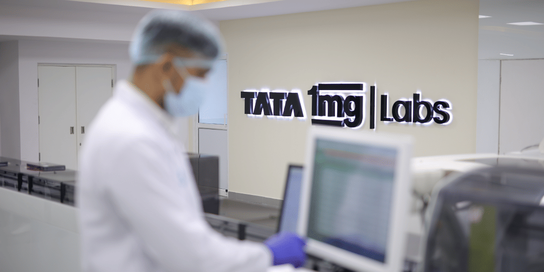
Tata 1mg Labs
NABL certified, ISO certified


Accredited
labs
Highly skilled
Phlebos
Verified
reports
Who will collect your samples?
Tata 1mg certified phlebotomists
Know more about lab
NABL certified, ISO certified
Test Information is reviewed by: Dr. Swati Gupta (Anand), MBBS, MD Lab Medicine and written by Dr. Lipika Khurana, BDS, PGDHHM . The test details are for information purpose only. Consult a doctor before taking any test. Last updated on: 23rd Dec 2025
Did you find information useful ?
Book a Fever Package (includes Malaria & Typhoid tests) Old
test at home near me
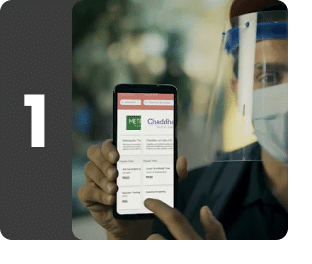
Easy online booking
Search for tests and packages, book a time and select address for seamless at-home lab tests.
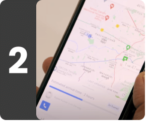
Live tracking of phlebotomist
Stay informed with live tracking of our phlebotomist's location for seamless sample collection.
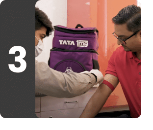
Safe collection
Our phlebotomists follow strict safety protocols to collect samples at home on time.
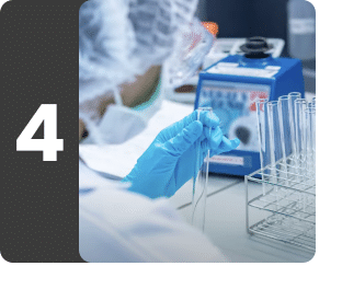
Sample received at lab
Your sample is bought to our laboratory for testing by our qualified experts.
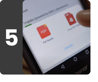
Quick reports, free follow up
Reports are sent to your email. A free doctor follow up is provided to understand the report better


Recommended for everyone
This package is designed with everyone’s overall health considerations in mind, offering assessments to address a wide range of wellness needs.
Package contains 18 tests
Report delivery
Standard time
12 hrs
For slots after 11 AM, report will be delivered in 24 hours.
Preparations
1
Urine sample must preferably be the midstream urine (part of urine that comes after first and before the last stream). Collect the urine sample in a sealed and sterile container provided by our sample collection professional. Make sure that the container doesn't come in contact with your skin. Please be informed that urine sample is a part of this package, you are required to submit all the samples that are a part of the package during the sample collection itself. Women are advised not to give the sample during the menstrual period unless prescribed.
Why is this package booked?
1
If you are suffering from chronic low grade fever
2
If you have signs or symptoms of typhoid (enteric) fever like headache, high fever, abdominal pain, diarrhea, weight loss or skin rashes
3
If you have signs or symptoms of malaria like fever with shaking chills, headaches
4
In suspected cases of pyrexia of unknown origin (PUO)
Who's behind your sample collection?


MEET YOUR PHLEBO

Certified & experienced professionals
Tata 1mg phlebotomists are DMLT / B.Sc MLT certified and have 1+ years of experience
Best in-class collections
Tata 1mg phlebotomists are trained for painless, single-prick hygienic sample collection

Comprehensive expertise
Other than sample collection, our phlebos are also skilled in first aid, ECG & BP monitoring
WHAT OUR CUSTOMERS ARE SAYING
Despite of very heavy rains, reached on time and did the work very professionally
~ Rahul Shrivastava
It didn't pain a 7 year old and hats off to the collector for being kind and extremely kid friendly
~ Khushnuma
Very nice person and soft spoken. I felt good talking to him even when I am in fever
~ Sourabh
New gloves new everything for each person, it's brilliant how meticulous and detailed your SOPs are.
~ Anita
My mother's veins are thin and slippery and hard to locate but Mohammed Usmani got it so fast, no pain
~ Rama Sampath
No pain at all while collecting blood sample. Fast, quick and properly trained staff.
~ Monalithatte
Sample collection agents was very professional and quick he was so organised with everything
~ Vaishali